Steven Beyea
Professor, Department of Physics and Atmospheric Science, Department of Diagnostic Radiology, School of Biomedical Engineering
Contact
Steven Beyea
Email: steven.beyea@dal.ca
Phone: 902-473-1868
Web:
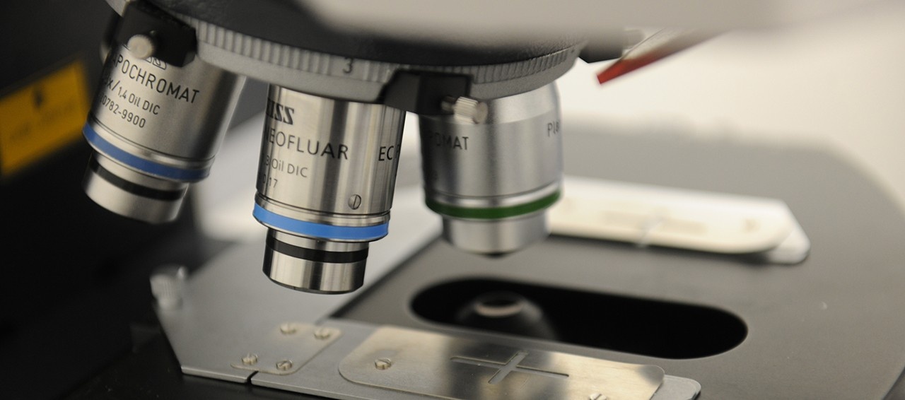
My laboratory focuses on the development, application and clinical/commercial translation of novel diagnostic imaging technologies. ╠²Research in the lab encompasses the development of acquisition techniques and analysis algorithms for parametric and functional mapping using clinical MRI and MEG (magnetoencephalography), as well as multimodal integration in both clinical and pre-clinical imaging.
Our Group
As the head of the Biomedical Translational Imaging Centre (BIOTIC), I take a highly interdisciplinary approach to medical imaging. Strategically located in HalifaxÔÇÖs largest hospitals, our infrastructure ranges from clinical 3T MRI and MEG, to pre-clinical MRI, PET, SPECT and CT. Students interested in interdisciplinary research projects, particularly in the areas of functional brain imaging and abdominal/pelvic cancer imaging, should contact me about ongoing research opportunities.
Projects
| Compressed Sensing Approaches to High Temporal Resolution Parametric Mapping: This project utilizes novel applications of image compression technology to the development of new techniques for obtaining high temporal resolution image data. This will be specifically applied across a range of MRI applications from functional neuroimaging to dynamic contrast enhanced MRI mapping of tumour pharmacokinetics. |
| Algorithms for Enhanced Reliability of fMRI and MEG in Pre-Surgical Functional Neuroimaging: While various functional neuroimaging techniques are widely used for pre-surgical mapping of critical functional zones in the brain, traditional analysis methods are not well suited to objectively generating reliable maps of brain function in individual patients. We are developing a new algorithm for an automated and data-driven approach to analyzing this data, and benchmarking it against cortical stimulation data obtained during brain surgery. |
| Novel MRI Pulse Sequences for Improved Quantification of Iron/Fat: Current clinical techniques for non-invasive mapping of liver iron/fat generally result in maps created using a priori assumptions about the fatty molecules and have iron context calculated from 3 to 6 data points. We are developing a new pulse sequence that will generate thousands of high temporal resolution data points for accurate mapping for fat and iron content, with no a priori assumptions on the molecular structure of the fat, with a goal of improved diagnosis of fatty liver disease. |
| Machine Learning Approaches to Improved Patient Stratification using MRI/MEG: Not all patients recover with the same ÔÇ£trajectoryÔÇØ nor respond to the same therapies. Using longitudinal data collected using MEG and/or MRI, we are using machine learning approaches to determining markers of brain function/connectivity that separate patients who should ideally be triaged into differing sub-populations in order to individualize treatment. This work is being applied to several clinical areas including brain injury and degeneration. |
╠²
Selected Publications
| D.P. McAllindon, C. Bowen, C. OÔÇÖGrady, S. Beyea╠² ÔÇ£An investigation of unbiased measures for automated thresholding and data quality estimation for pre-surgical fMRIÔÇØ╠²Proceedings of the╠²Joint Annual Meeting of the International Society for Magnetic Resonance in Medicine and the European Society for Magnetic Resonance in Medicine & Biology, Paris, France (2018). |
| S. Patterson, P. Lee, C. Bowen, J. Merrimen, C. Wang, S. Beyea, S. Clarke╠² ÔÇ£Machine Learning & Prostate Cancer Detection: Role of Individual Contrasts and Comparison with PI-RADSÔÇØ╠²Proceedings of the╠²Joint Annual Meeting of the International Society for Magnetic Resonance in Medicine and the European Society for Magnetic Resonance in Medicine & Biology, Paris, France (2018). |
| J.A. Rioux, N. Murtha, C.V. Bowen, S.E. Clarke, S.D. Beyea ÔÇ£Reconstruction of Accelerated DCE-MRI Guided by Image Quality MetricsÔÇØ Proceedings of the Joint Annual Meeting of the International Society for Magnetic Resonance in Medicine and the European Society for Magnetic Resonance in Medicine & Biology, Paris, France (2018). |
| Petley L, Bardouille T, Chiasson D, Froese P, Patterson S, Newman A, Omisade A, Beyea S. Brain Inj. 2018;32(4):464-473. ╠² |
| Rioux JA, Beyea SD, Bowen CV. MAGMA. 2017 Feb;30(1):41-55. ╠² |
| Stevens T, Bardouille T, Stroink G, Boe S, Patterson S, Beyea S. J Neurosci Methods. 2016 Jun 15;266:21-31. ╠² |
| Stevens MT, Clarke DB, Stroink G, Beyea SD, D'Arcy RC. J Neurol Neurosurg Psychiatry. 2016 Mar;87(3):267-74. |
| T. Bardouille, T. Stevens, S. Boe , S.D. Beyea ÔÇ£Enhanced Clinical MEG Pre-Surgical Functional Mapping: Reliability & Spatiotemporal ClusteringÔÇØ Proceedings of the 20th Annual Meeting of the Organization for Human Brain Mapping, Hamburg, Germany (2014) |
| M.T.R. Stevens, R.C.N. DÔÇÖArcy, G. Stroink, D.B. Clarke & S.D. Beyea "Thresholding in fMRI Studies: At the Cost of Reliability?" J. Neuro. Methods 219, 312-323 (2013) |
| D. Holland, C. Liu, E. Mazerolle, X. Song, T. Stevens, C. V. Bowen, A. Sederman, L. Gladden, S. D. Beyea "Compressed Sensing Reconstruction Improves Sensitivity of Variable Density Spiral fMRI " Magn. Reson. Med., 70(6), 1634-43 (2013) |
| J.M. Bray, M. Filiaggi, C.V. Bowen and S.D. Beyea ÔÇ£Degradation & Drug Release in Calcium Polyphosphate Bioceramics: An MRI-Based CharacterizationÔÇØ, Acta Biomaterialia 8, 3821-3831 (2012) |
| S.R. McWhinney, E.L. Mazerolle, J.R. Gawryluk, S.D. Beyea & R.C.N. D'Arcy "Comparing gray and white matter fMRI activation using asymmetric spin echo spiral" J. Neurosci. Methods 209, 351-356 (2012) |
| K.D. Brewer, J.A. Rioux, M. Klassen, C.V. Bowen and S.D. Beyea ÔÇ£Signal Displacement in Spiral-In Acquisitions: Simulations & Implications for Imaging in SFG RegionsÔÇØ Magnetic Resonance Imaging 30, 753-763 (2012) |
| J.A. Rioux, K.D. Brewer, S.D. Beyea, C.V. Bowen "Quantification of Superparamagnetic Iron Oxide with Large Dynamic Range using turboSPI" J. Magn. Reson., 216, 152-160 (2012) |